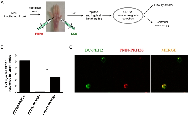
Figure 9. Co-injection of PMNs with endocytosed bacteria and DC in subcutaneous tissue gives rise to the detection of DC carrying PMN-derived material in the draining lymph node.
(A) Schematic representation of experiments in which bone marrow-derived DC and PMNs that had been pre-exposed to E. coli and washed were injected in separate syringes into the foot pads of mice. Both leukocyte types were differentially labelled with fluorescence dyes (PKH26 for PMNs and PKH2 for DC). (B) Calculations of the percentages of injected DC recovered as CD11c+ immunomagnetically selected cells from popliteal lymph nodes measuring double or single positive fluorescent events as indicated by flow cytometry. (C) Representative confocal images of cytospins made with the CD11c-immunoselected cells from popliteal lymph nodes. Experiments are representative from at least two similarly performed with three mice per group each. Asterisks indicate statistical significance p<0.01 in student's t test.

