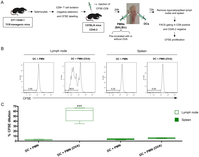
Figure 10. Cross-presentation of antigens taken by DC from PMNs take place in vivo.
(A) Schematic representation of experiments in which splenocytes from OT1 CD45.1 TCR-transgenic mice were subjected to CD8+ T cell isolation by immunomagnetic negative selection. OT-1 Cells were labeled with CFSE and 2×10⁶ were injected intravenously into C57BL/6 mice. 24 h later mice received injections of PMNs and DC (H-2b) in the footpad. Immunomagnetically isolated PMN were from BALB/c mice (H-2d) and were pre-incubated or not with OVA for two hours and extensively washed. PMNs and DC were injected with different syringes in the footpads of the mice which had been transferred with CFSE-labelled OT-1 cells 24 hours before. 72 h later proliferation of OT1 CD45.1+ cells was assessed by flow cytometry in draining lymph node and spleen (B) Representative histograms of CFSE dilution seen in lymph nodes and spleen. (C) Summary data of CFSE expression from lymph nodes and spleens in 3 experiments performed as described in A. When indicated PMN were pre-incubated or not with OVA. Asterisks indicate statistical significance p<0.001 in Mann-Whitney U test.

