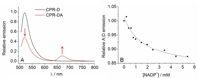
Figure 2. Coenzyme binding causes CPR to close.
(A) Fluorescence emission spectra of CPR-D (black line) excited at 495 nm and CPR-DA excited at 495 nm (red line). The introduction of an acceptor fluorophore causes a decrease in donor emission and an increase in acceptor emission when the donor is excited (red arrows), which is demonstrative of FRET. The same concentration of donor fluorophore is present in both spectra. (B) Effect of titrating NADP+ on the relative change in FRET efficiency (expressed as variation in A∶D ratio). The solid line shows the fit to Equation 1. Conditions: 50 mM potassium phosphate pH 7, 25°C, 0.3 µM CPR-D, 0.6 µM CPR-DA.

