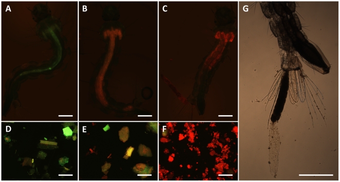
Figure 8. Detection of C14 porphyrin in exposed larvae and PFP particles.
Ae. aegypti larvae were exposed to the photosensitizer at various conditions of food incubation. In particular, A, D: control; B, E: filtration eluate (experimental group B; see Fig. 6); C, F: typical example of larva and food particles from the other porphyrin treatments (experimental groups A, C and D; see Fig. 6). Larval food, either fresh or pre-incubated with porphyrin, was added in the trays at the same time of the introduction of the larvae, in all the groups. G: typical extrusion of the gut lining and content observed in C14 porphyrin-treated larvae (also visible in C). Scale bars represent 500 µm.

