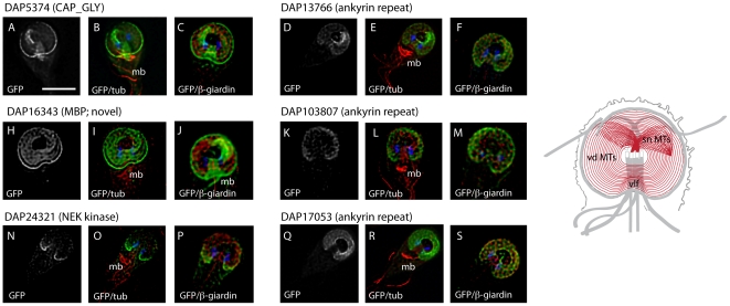
Figure 3. Novel disc-associated proteins associated with the entire disc.
Six new DAPs localize to the entire ventral disc spiral in a manner similar to the previously described microribbon-associated proteins (see Figure 2 and Figure S3) as visualized by C-terminal GFP tagging (grey or green) and either anti-alpha-tubulin immunostaining of the MT cytoskeleton (red), or anti-beta-giardin immunostaining of the ventral disc microribbons (red). The two nuclei are visible with DAPI staining (blue). One putatively microtubule-associated DAP possesses a CAP-Gly motif (A,B,C). Another disc-localizing DAP is the possibly mis-named median body protein (MBP, H,I,J). Three ventral disc-localizing DAPs have ankyrin repeat domains (D–F, K–M, and Q–S). Note the absence of GFP localization in the ventrolateral flange (vlf) area in DAP17053 (Q–S). Finally, one disc-localizing DAP is a Nek kinase that has a greater localization to the posterior region of the ventral disc (N–P). Scale = 5 µm. Schematic shows areas of GFP localization in red.

