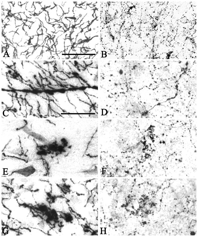
Figure 2.
In the youngest case investigated (Case #1), AChE activity (A) and ChAT immunoreactivity (B) were present within cholinergic axons homogenously thin in diameter. Immunohistochemistry for ChAT visualized small varicosities within axons. In brains from older cases and AD brains, one or more of a number of abnormalities were seen in both AChE (C, E and G) and ChAT (D, F, H) stained material. These included thickened axons (C and D) and swollen and ballooned varicosities at axonal endings (E–H), which often occurred in a chandelier arrangement. Scale bar in A is 100 μm and also applies to B, and the scale bar in C is 50 μm and also applies to D–H.

