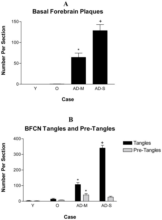
Figure 4.
The number of basal forebrain plaques, tangles and pre-tangles within cholinergic neurons in each section. Plaques were absent from the basal forebrain of young individuals. They were seen in old individuals in moderate numbers. A significant increase in the number of plaques was seen in pathologically mild AD and another significant increase in pathologically severe AD. The density of pre-tangles was higher in pathologically mild AD when compared with non-demented old cases. The density of tangles showed a further significant increase in the cholinergic neurons in pathologically severe AD. Quantitative analysis was carried out using all of the cases in Table 1 except those identified by an asterisk. A. *p<0.001 compared with non-demented old cases, +p<0.001 compared with pathologically mild cases. B. *p<0.001 compared with non-demented old cases, +p<0.001 compared with pathologically mild cases. Y – Non-Demented Young, O – Non-Demented Old, AD-M, Pathologically Mild AD, AD-S – Pathologically Severe AD.

