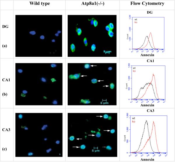
Fig. 2. Spontaneous PS-externalization in the hippocampus of the Atp8a1(-/-) but not the wild type mice.
(a-c) Alexafluor488-Annexin V (green) and HOECHST33342 (blue) staining of cells from the neuronal layers in DG, CA1, and CA3 of control and Atp8a1(-/-) mice. Merged images of annexin V(+) cells were observed in the cells from the Atp8a1(-/-) mice. White arrows show non-apoptotic but annexin V(+) cells. (Flow Cytometry, Right panel) From three experiments, the increases in mean fluorescence were 3.6-fold (DG), 3.9-fold (CA1), and 10.7-fold (CA3) in the Atp8a1(-/-) samples (red traces).

