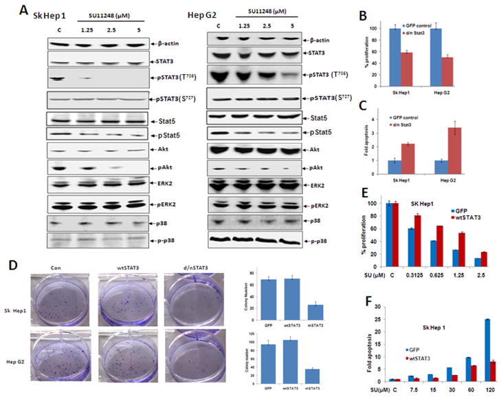
Fig. 5.
STAT3 is implicated in the mechanism of sunitinib-induced suppression of HCC growth. (A) Sk Hep1 or Hep G2 cells were seeded in 60mm petri dishes at a concentration of 2×10⁶/dish and then treated with sunitinib at the indicated concentrations for 24 hr. Cells were then lysed and proteins extracted, quantified, and analyzed by Western blotting with antibodies specific for the indicated proteins, and β-actin as an internal control. (B-D) Sk Hep 1 or Hep G2 cells were transfected with wtSTAT3 and dnSTAT3 and maintained overnight. Some of the cells transfected with dnSTAT3 were replated in 96-well plates at a density of 5×10⁴/well the following day and used for evaluation of proliferation (B) or suppression of apoptosis (C). In addition, some of the cells transfected with wtSTAT3 and dnSTAT3 were replated at a density of 300 cells/well in 6-well plates for colony forming assays (D). n=3, error bars represent mean +/- S.D.

