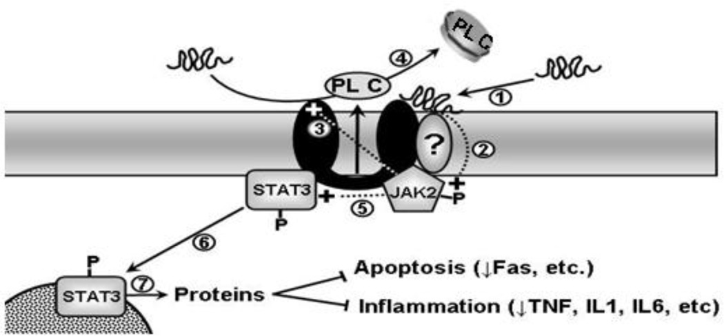
Figure 1. Scheme for the apoA-I/ABCA1/JAK2/STAT3 pathway.
Lipid-poor apoA-I or its mimetic peptides bind directly to ABCA1 or a closely associated protein (step 1) that rapidly stimulates JAK2 autophosphorylation (step 2). The activated JAK2 then has two distinct effects on the cells. First, it recruit and/or dissociate ABCA1 partner proteins and causes confirmation change of ABCA1, thus enhancing apoA-I binding to ABCA1 (step 3) and lipid removal from the cells (step 4). Second, it generates STAT3 docking sites and activates STAT3 (step 5), which promotes translocation of STAT3 to the nucleus (step 6) where it regulates transcription of proteins that repress inflammatory processes (step 7). P: Phosphorylated.

