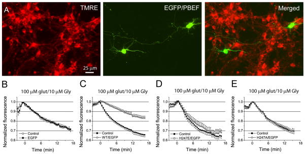
Fig 6. Overexpression of PBEF with enzymatic activity reduces mitochondrial membrane (MMP) depolarization induced by excitotoxic glutamate stimulation.
(A) Fluorescence images of EGFP-expressing and TMRE-labeled neurons. (B–E) Comparison of time courses of TMRE fluorescence changes in neurons expressing EGFP alone (B), and neurons coexpressing EGFP and WT hPBEF (C), EGFP and H247E (D) and H247A (E) mutants with non-transfected neurons after glutamate stimulation. Notice PBEF-expressing neurons have much slower rate of MMP depolarization than neurons expressing PBEF mutants, EGFP alone, and non-transfected neurons. Data was normalized to control value before glutamate application. All data was collected from 3–4 preparations of primary cultured neurons.

