
Figure 3. The crystal structures of MACPF family proteins showing a CDC domain 3 like structure.
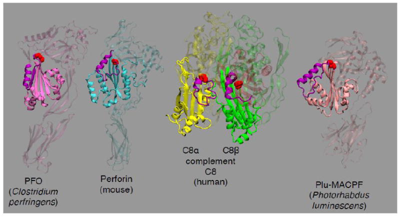
The crystal structures of the MACPF proteins mouse perforin (PDB ID: 3NSJ) [88], complement C8 (PDB ID: 3OJY) [120] and the Photorhabdus luminescens Plu-MACPF (PDB ID: 2QP2) [89] are shown with that of PFO [32]. In each MACPF structure the PFO-like domain 3-like fold is highlighted. Shown in purple ribbon structure in each MACPF structure is the equivalent structure to the PFO α1-β5 loop that rotates away from β4. Shown in red spacefilled atoms is the conserved twin glycine motif of each protein that is present at the junction between β4 and β5, which is conserved in all know CDCs [46].