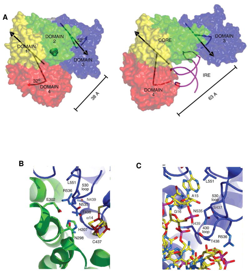
Figure 3. Crystal structure of IRP1 as c-aconitase and complexes with IRE-RNA.
A. Differences in protein domain positions between c-aconitase and IRP1:IRE-RNA complex. Left: FeS-apo-IRP; Right: IRE-RNA/IRP. RNA-magenta; protein- blue, green, red, yellow. B. Close up of the protein–RNA contacts at the RNA triloop; C. Close-up of the protein–RNA contacts at the RNA bulge C8. (From ref. 12, with permission).

