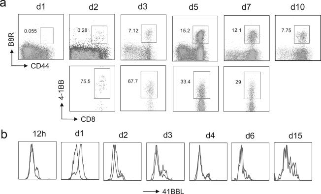
Figure 2.
4-1BB and 4-1BBL could be detected on VACV-specific CD8 T cells and DC respectively following infection. a-b) WT mice were infected i.p with VACV-WR (2 × 10⁵ PFU/mouse). a) On indicated days post-infection splenocytes were harvested and stained for CD8, CD44, B8R-tetramer, and 4-1BB. Top panel, Percentage of CD44-high expressing B8R-specific CD8 T cells. Bottom panel, Percentage of 4-1BB+ cells gating on CD8+ CD44-high B8R-tetramer positive cells. Quadrant settings based on isotype controls. b) On indicated days post-infection splenocytes were harvested and stained for CD8, CD11c, CD11b, and 4-1BBL. Representative histogram of 4-1BBL expression gated on CD8+CD11c+ cells.

