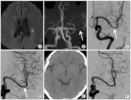
Fig. 1.
A 70-year-old man presented with minor stroke with national Institutes of Health Stroke scales 2. A : Diffusion-weighted image shows a focal acute infarction in the left corona radiata. B and C : Magnetic resonance angiography (B) and left internal carotid artery angiography (C) images show a focal severe stenosis of the distal first segment of the left middle cerebral artery. D : Immediate post-stenting angiogram shows complete recanalization of the stenotic segment with normalized luminal diameter. E : Post-procedural computed tomography shows no intracranial hemorrhage. F : Angiogram obtained 19 months after the stenting reveals no significant in-stent restenosis.

