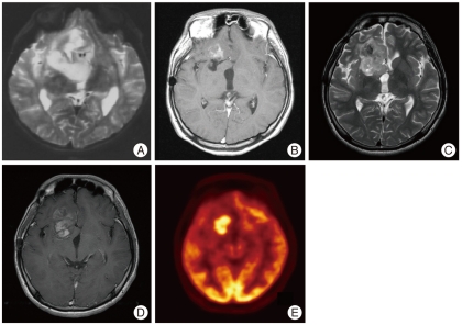
Fig. 1.
A : Preoperative T2-weighted image from the surgical specimen at the first operation demonstrates heterogeneous signal intensity in a right frontal mass. B : Contrast-enhanced T1-weighted image shows subtle peripheral enhancement 13 years after the first operation. C : T2-weighted image taken 15 years after the first operation demonstrates high signal intensity mass lesion in the right frontal lobe. D : Contrast-enhanced T1-weighted image taken 15 years after the first operation shows marked enhancement. E : 18F-FDG brain positron emission tomography shows a hypermetabolic lesion in the right inferior frontal lobe.

