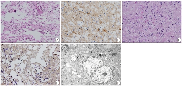
Fig. 2.
A : H&E staining shows round to oval, small uniform cells from the specimen taken at the first operation (Magnification; ×200, H&E stain). B : Immunostaining for synaptophysin demonstrates a strong positivity in the specimen taken at the first operation (Magnification; ×400, synaptophysin immunochemical stain). C : Small round cells with large clear cytoplasms and round nuclei (Magnification; ×400, H&E stain). D : A strong positivity for synaptophysin is seen (Magnification; ×400, synaptophysin immunochemical stain). E : Electron microscope shows that the cytoplasmic organelles are sparse and include a few strands of RER cisternae, dense bodies and glycogen particles (Magnification; ×7,000, electron microscopy).

