Fig.1.
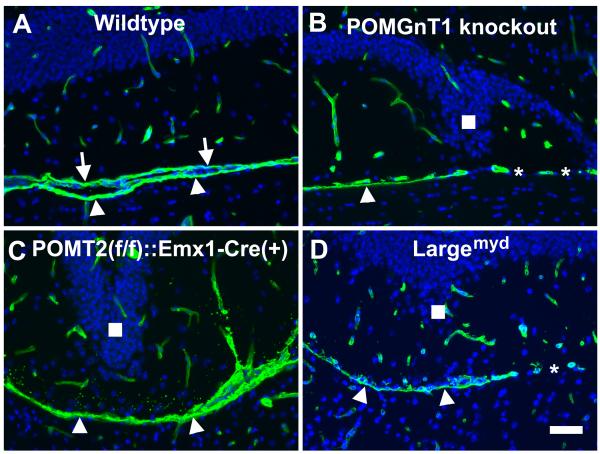
Laminin immunofluorescence staining suggests disruption of the pial basement membrane in the dentate gyrus of CMD mouse models.
Coronal sections of adult forebrain were immunostained with anti-laminin (green fluorescence) and counterstained with DAPI (blue fluorescence). (A) Wild type. Laminin immunofluorescence was observed in blood vessels and pial basement membrane. The pial basement membrane of the dentate gyrus (arrows) and the midbrain (arrowheads) were clearly discerned. (B) POMGnT1 knockout. In some regions only a single pial basement membrane, presumably of the midbrain, was observed (arrowhead). There were regions devoid of any pial basement membrane separating the dentate gyrus and the midbrain (asterisks). (C) POMT2f/f;Emx1-Cre(+). A single pial basement membrane is observed between the dentate gyrus and the midbrain (arrowheads). (D) Largemyd mice. Regions of single pial basement membrane (arrowheads) and regions with no pial basement membrane were observed. Scale bar in D: 50 μm.

