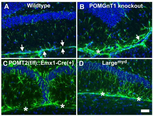
Fig.3.
Reactive astrogliosis was present at the dentate gyrus of the knockout animals.
Coronal sections of adult brain were immunofluorescence stained with anti-GFAP (green fluorescence) and counterstained with DAPI counter staining (blue fluorescence). (A) Wildtype. GFAP immunofluorescence was observed at two continuous glia limitans separating the dentate gyrus and the midbrain (arrows). GFAP-positive astrocytes were sporadically positioned within the lower blade of the dentate. (B) POMGnT1 knockout. Glia limitans were observed (arrow) but discontinuous (asterisk indicates breaks). GFAP-positive astrocytes with increased size were frequently observed within the lower blade. (C) POMT2f/f;Emx1-Cre(+). Breaks in glia limitans (asterisks) were frequently observed. GFAP-positive astrocytes were increased over the wildtype. (D) Largemyd mice. Breaks in glia limitans were observed (asterisks) and increased GFAP-immunofluorescence was apparent. Scale bar in D: 50 μm.

