
Fig. XX.1. Effects of intermittent and constant high glucose on ER stress and MCP-1 secretion.
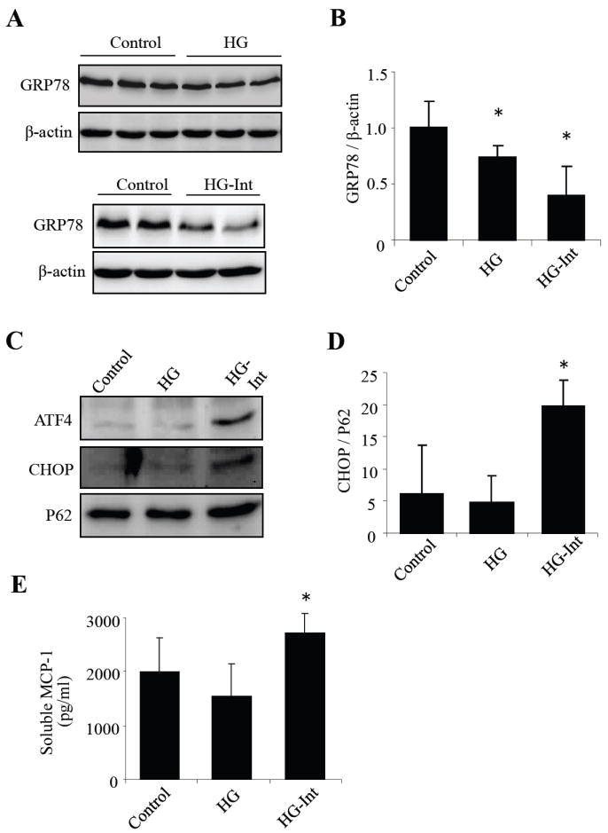
A: GRP78 expression, increases MCP-1 level and activates ER stress-mediated apoptosis in human retinal pericytes. HRP were exposed to stable high glucose (HG, 25 mM), intermittent high glucose (HG-Int, intermittent exposure to 25 mM glucose at 48h intervals) or normal glucose (5 mM) for 8 days. (A-B) Expression of GRP78 was detected by Western blot analysis in whole cell lysates. (C) Protein level of GRP78 was quantified by densitometry (mean ± SD, n=3). (D) Expression of ATF4 and CHOP was determined by Western blot analysis in nuclear extracts. (F) Protein level of CHOP was semiquantified by densitometry (mean ± SD, n=3). (F) Soluble MCP-1 secreted into the medium was measured using ELISA (mean ± SD, n=3). *P<0.05 vs. control.