Figure 2.
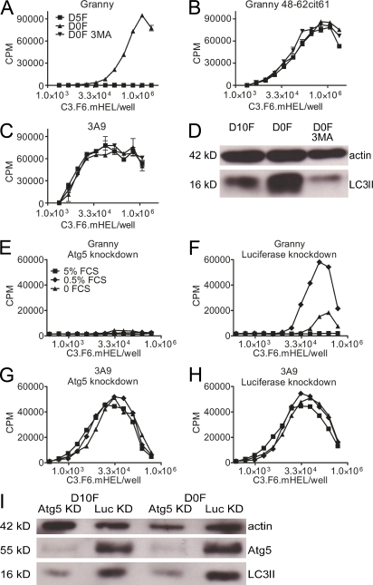
C3.F6.mHEL cells present citrullinated peptides after serum starvation–induced autophagy. (A–C) T cell responses to C3.F6.mHEL cultured in DME supplemented with 5% FCS (D5F), no serum (DOF), or no serum in the presence of 10 mM 3MA (D0F 3MA) for 4 h; the culture medium was then changed to 5% FCS, and the T cell hybridomas Granny (A and B) or 3A9 (C) were added. (D) Western blot of actin and LC3II in C3.F6.mHEL cultured in D10F, D0F, and D0F with 3MA. Data are representative of three independent experiments; each data point represents duplicate wells. Targeted knockdown of Atg5 expression inhibited citrullination of HEL peptides. (E–H) Responses of T cells to C3.F6.mHEL cells expressing either shRNA targeting Atg5 expression (E and G) or luciferase expression (F and H). Cells were cultured in media supplemented with 5% FCS, 0.5% FCS, or no FCS for 4 h and then replaced with media supplemented with 5% FCS. (I) Western blot of actin, Atg5 levels, and LC3II in the treated C3.F6.mHEL cells shows that Atg5-deficient cells had reduced levels of LC3II conversion after serum starvation. Data are representative of three independent experiments. Error bars indicate SEM.

