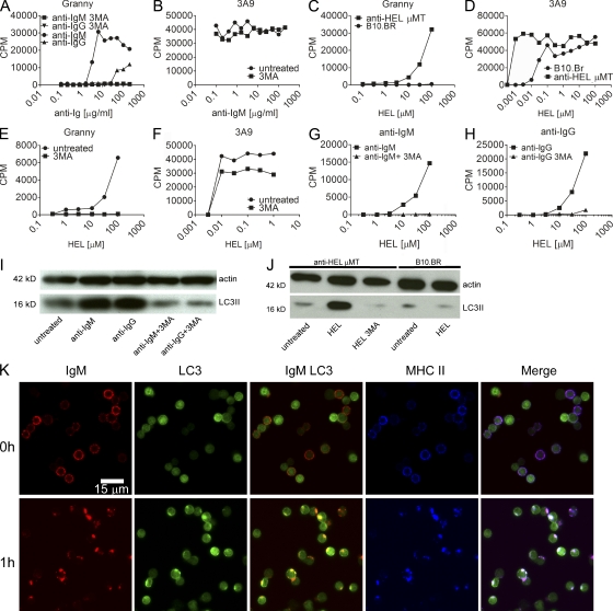
Figure 5.
BCR engagement induces presentation of citrullinated peptides by primary B cells. (A and B) Presentation by CD19+ cells isolated from spleens of mHEL mice. (A) Effects of various concentrations of anti-IgM or anti-Ig on Granny ± 3MA. (B) The same as in A but on 3A9 ± 3MA. (C) Presentation of HEL by CD19+ cells from anti-HEL transgenic mice or B10.BR mice to Granny. (D) The same as in C but to 3A9. (E) The effect of 3MA on presentation by CD19+ cells from anti-HEL transgenic mice after processing HEL to Granny. (F) The same as in E but to 3A9. (G) Presentation of HEL by B cells from B10.BR mice to Granny after treatment with 40 µg/ml anti-IgM ± 3MA. (H) The same as in G but treating the cells with anti-IgG ± 3MA. (I and J) Western blots of actin and LC3II from mHEL B cells cultured overnight with anti-Ig (I) or anti-HEL B cells cultured with 30 µM HEL ± 3MA (J). (K) B cells from GFP-LC3 mice were labeled on ice with anti-IgM and fixed immediately or after 1 h at 37°C and stained to label MHC class II. All data are representative of at least two independent experiments.

