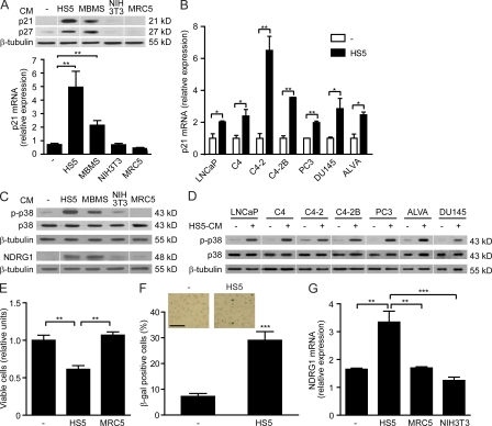
Figure 1.
CM of bone stromal cells induces senescence in prostate cancer cells. (A) The prostate cancer cell line PC3 mm was cultured in the presence of CM from human bone stromal cells (HS5), mouse BM stroma (MBMS), mouse embryonic fibroblasts (NIH3T3), or human lung fibroblasts (MRC5) or serum-free medium (−). Expression of p21, p27, and β-tubulin was examined by qRT-PCR (n = 3) and Western blot. (B) The prostate tumor cell lines LNCaP, C4, C4-2, C4-2B, PC3, DU145, and ALVA were cultured in the presence of CM of HS5 or serum-free medium (−), and the expression of p21 was examined by qRT-PCR (n = 3). (C) PC3 mm was cultured in the presence of CM of HS5, MBMS, NIH3T3, or MRC5 or serum-free medium (−), and the activation of p38 (phosphorylated p38, p-p38; total p38, p38) and the expression of NDRG1 and β-tubulin were examined by Western blot. (D) LNCaP, C4, C4-2, C4-2B, PC3, ALVA, and DU145 were cultured in the presence or absence of CM of HS5, and the expression of p-p38, p38, and β-tubulin was examined by Western blot. (E) PC3 mm was cultured in the presence or absence of CM of HS5 or MRC5 for 48 h, and cell viability was measured by the MTT assay (n = 3). (F) PC3 mm was cultured in the presence or absence of HS5-CM for 48 h, and SA–β-gal staining (blue-green) was performed (n = 4). The inset shows representative images of three independent experiments. Bar, 100 µm. (G) PC3 mm was cultured in the presence of CM of HS5, MRC5, or NIH3T3 or serum-free medium (−), and the expression of NDRG1 was measured by qRT-PCR (n = 3). *, P < 0.05; **, P < 0.01; ***, P < 0.001. All experiments were performed three times independently, and representative data are shown. Results are shown as mean ± SEM.

