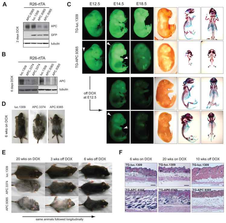
Figure 5. APC loss disrupts embryonic and postnatal development.
(A) Western blot of whole protein lysates from E10.5 embryos on DOX for 2 days and(B) E14.5 embryos on DOX for 6 days.
(C) GFP and brightfield images of embryos treated with DOX from E8.5 or pulsed with DOX from E8.5–E12.5. Arrows indicate fluid accumulation along the dorsal ridge and defects in limb and digit development at E12.5 and E14.5. Alcian blue and Alizarin red stained skeletons from E18.5 embryos.
(D) Representative photographs of TG-luc.1309/R26-rtTA, TG-APC.3374/R26-rtTA and TG-APC.9365/R26-rtTA double transgenic mice on DOX for 6 weeks.
(E). Representative photographs of Luc.1309, APC.3374 and APC.9365 treated with DOX for 20 weeks and then removed from DOX-treatment for 6 weeks.
(F) H&E sections of skin taken from Luc.1309/R26-rtTA and TG-APC.9365/R26-rtTA double transgenic mice treated with DOX for 6 weeks, 20 weeks and 20 weeks on DOX/6 weeks off DOX as indicated. Scale bars are 100μm.
See also Figure S5.

