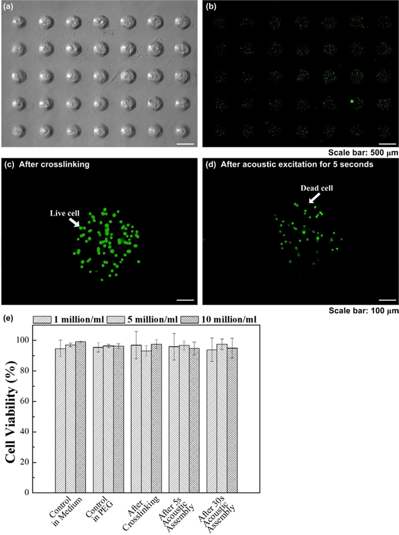
Figure 5. Viability of cells encapsulated in microgels during the acoustic assembly process.
(a) Fabricated cell-encapsulating microgels and (b) corresponding fluorescent images of live/dead staining after crosslinking. Cell viability in individual microgels after (c) crosslinking and (d) after acoustic excitation for 5 seconds. (e) Quantification of cell viability in medium, in PEG, after crosslinking and after acoustic excitation (5 seconds and 30 seconds).

