
Figure 1.
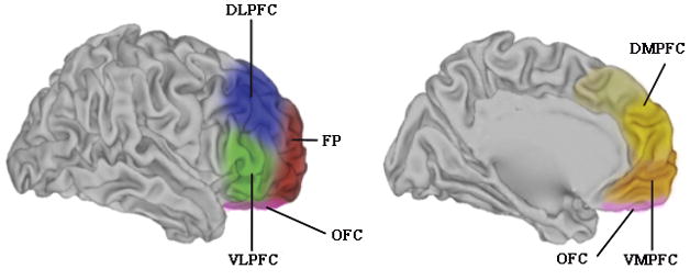
General regions of the PFC in humans. The colored regions represent rough approximations of the broad zones of PFC. In both the lateral view (left) and the medial view (right), the regions are overlaid on a “partially inflated” hemisphere that allows clear visualization of sulci. Abbreviations: DLPFC dorsolateral prefrontal cortex; VLPFC ventrolateral prefrontal cortex; FP frontopolar cortex; OFC orbitofrontal cortex, DMPFC dorsomedial prefrontal cortex; VMPFC ventromedial prefrontal cortex. Figure adapted with permission from mindblog.dericbownds.net.