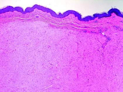
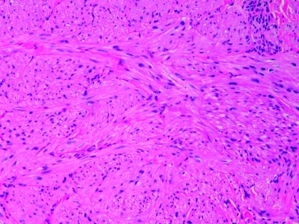
Figures 3A and 3B.
Microscopic features. A) Scanning magnification revealing poorly circumscribed nodules of eosinophilic spindle cells filling the dermis and extending into the subcutis (hematoxylin and eosin, original magnification 40x); B) higher magnification demonstrating fascicles of benign smooth muscle bundles without necrosis or nuclear atypia (hematoxylin and eosin, original magnification 100x)


