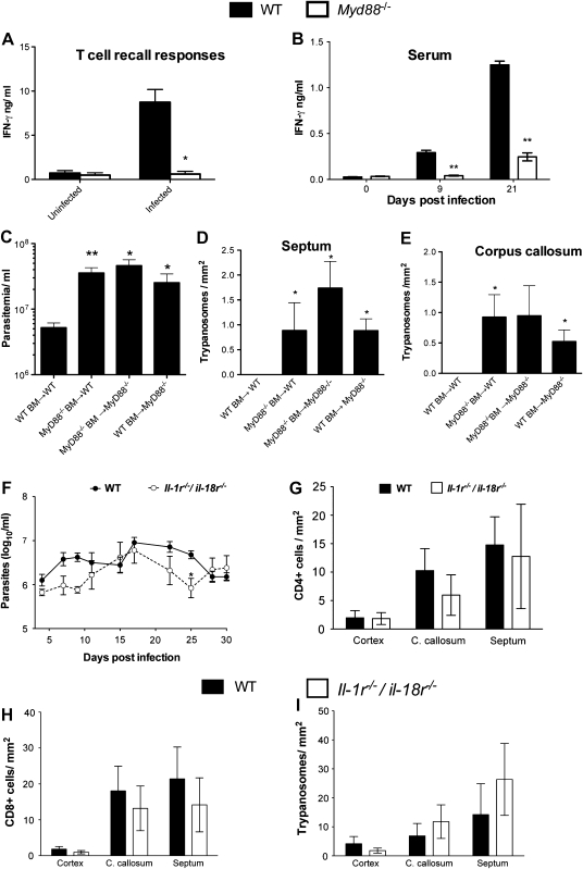
Figure 3.
MyD88 regulates the recall T-cell responses in the periphery, serum levels of interferon (IFN) γ, and parasitotoxic responses in the brain parenchyma. A, T-cell recall responses. Purified splenic T cells from wild-type (WT) or Myd88−/− mice infected with Trypanosoma brucei for 25 days or not infected were cocultured with syngeneic bone marrow (BM)–derived dendritic cells in the presence of T. brucei brucei lysates. IFN-γ was assayed by enzyme-linked immunosorbent assay in culture supernatants 72 hours after incubation. The means ± standard error of measurement (SEM) from triplicate cultures per condition are depicted. The experiments were performed twice with similar results. *P < .05 (unpaired t test; significant differences between WT and Myd88−/− cells). B, Serum IFN-γ levels after T. brucei brucei infection. Each bar represents mean ± SEM of values obtained from 3 or 4 animals per group and time point. **P < .01 (unpaired t test; significant differences compared with uninfected animals). C–E, Parasitemia (C) and trypanosome density in the septum (D) and corpus callosum (E) of radiation chimeric mice as measured 15 days post infection with T. brucei brucei. Mean parasitemia and parasite density (± SEM) in brain sections of 5 mice per group. Differences compared with WT BM→WT sham chimeras are significant (*P < .05, **P < .01; unpaired t test). F, Levels of parasitemia in infected WT and Il-1r−/−/Il-18r−/− mice. Each point represents mean log10 parasites per milliliter (± SEM) obtained from 5–9 animals per group. *P < .05 (2-way analysis of variance; significant differences compared with infected WT animals). G–I, Mean numbers (± SEM) of CD4+ T cells (G), CD8+ T cells (H), and T. brucei brucei (I) per square millimeter in the cerebral cortex, corpus callosum (C. callosum), and septal nuclei of infected WT and Il-1r−/−/Il-18r−/− mice at 30 days post infection (4 animals per group).

