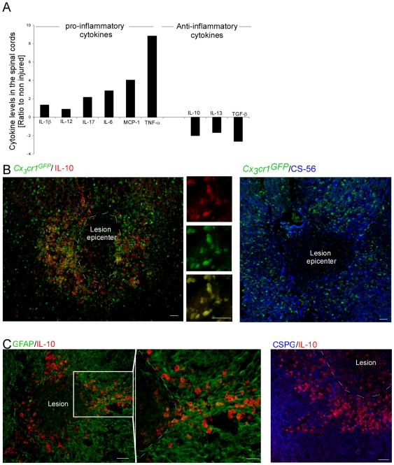
Figure 1. Resolving macrophages are restricted to a region enriched with glial scar matrix.
(A) Luminex analysis of the cytokine profile at the injured spinal cord. The results are presented as the ratio of expression levels relative to non-injured animals. Pooled samples (n = 3) were analyzed. The results are presented as change relative to the non-injured tissue. One representative experiment is shown out of two repetitions, each conducted at three different time points during the first week post injury (d1,3,7). The same tendency was observed for each time point tested, and in each repetition. The injury skews the local environment towards a pro-inflammatory milieu. (B) Spinal cord sections of injured [Cx3cr1 GFP/+>wt] BM chimeras, isolated at day 7 post injury, were co-stained for the infiltrating monocytes by GFP (green), and for the anti-inflammatory cytokine, IL-10 (red), or the glial scar matrix component, CSPG (CS-56; blue). (C) Injured spinal cord sections, isolated at day 7 post injury, co-stained to reveal the glial scar (astrocytes appear in green, and CSPG matrix protein in blue) and IL-10 (red), showing that resolving macrophages (rMΦ) are restricted to the CSPG-enriched area. Scale bar; 50 µm.

