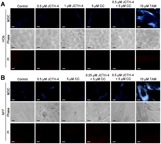
Figure 12. JCTH-4 and CC do not induce autophagy in Hob and NFF cells.
MDC staining was used to detect the presence of autophagic vacuoles in (A) HOb and (B) NFF cells after 72 hours of treatment with JCTH-4, CC, and solvent control (Me2SO) at the indicated concentrations. Bright blue punctate marks are indicative of autophagic vacuoles. Corresponding phase and PI micrographs are shown below the MDC images. Scale bar = 15 µm. All images are representative of 3 independent experiments.

