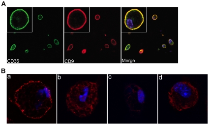
Figure 2. Co-localization of CD9 and CD36 on macrophage plasma membrane.
(A) Confocal Microscopy. Mouse peritoneal macrophages were seeded on glass coverslips, fixed in 4% formaldehyde, and then incubated with FITC-conjugated anti-CD36 IgA (left panel; green fluorescence) or unlabeled rabbit anti-CD9 IgG followed by Alexa-594 conjugated goat anti-rabbit IgG (middle panel; red fluorescence). Cells were also incubated with DAPI (Blue) to detect nuclei. Confocal images were obtained at (63×); insets show (6×63×). Right panel shows merged images. (B) Proximity Ligation Cross-linking Assay. Macrophages from wild type (a) or cd36 null (b) mice were incubated with rat anti-CD36 monoclonal IgG and rabbit anti-CD9 antibody and then species specific DNA-conjugated secondary antibodies. Specific oligonucleotides were then added, ligated and amplified using complementary fluorescent probes. Fluorescent dots represent cross-linked antibodies. In panels c and d, wild type macrophages were incubated with rabbit anti-CD36 IgG, but with anti-CD31 (c) or anti-CD40 (d) as negative controls.

