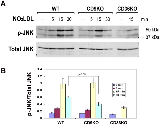
Figure 4. OxLDL induced JNK phosphorylation is reduced in cd9 null macrophages.
(A) Peritoneal macrophages from wild type, cd9 null and cd36 null mice were stimulated by oxidized LDL (50 µg/ml) for timed points from 0–30 minutes. Cells were then lysed and analyzed by western blot with antibodies to phosphor-JNK (top) or total JNK (bottom). (B) Blots from (A) were scanned and band densities quantified using NIH Image-J software. The ratios of p-JNK/total JNK are indicated, each group represents the mean of 3 individual samples.

