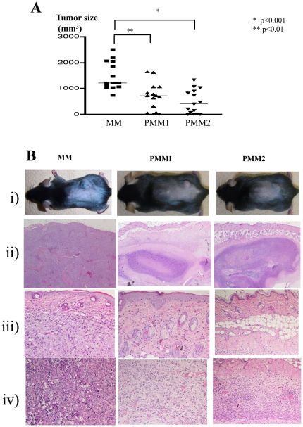
Figure 1. Tumor size, clinical phenotype, and histological findings.
A. Tumor size at the end of observation period: 4 weeks after B16 melanoma implantation. PBS-treated control mice (MM) developed a large tumor. Treatment with P. acnes led to a significant decrease of tumor size in both PMM1 (p<0.01) and PMM2 (p<0.001). B. i) Clinical picture of the tumors implanted on the dorsal skin. ii) Massive melanoma cell proliferation in the subcutis with invasion of the surrounding muscular tissues in MM. By contrast, PPM1 and PMM2 displayed subcutaneous granuloma formation (×40). iii) Close up of the upper dermis (×200). iv) Close up of the muscular tissue levels (×200). In MM mice, melanoma cell growth was observed without inflammatory cell infiltration. In PMM1 mice, melanoma cells were detected around the muscular lesion, but melanoma cells were almost undetectable, and abundant mononuclear cell infiltration was found in the PMM2 mice.

