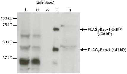
Figure 3. Western blot analysis of F2A efficiency at the Bapx1 locus.
Cell lysates from ten E12.5 FLAG3-Bapx1FE embryos were affinity purified using anti-FLAG antibody-conjugated beads. Protein fractions from total lysate, first and last wash, eluate and boiled beads were subjected to SDS-PAGE. Immunoblotting by anti-Bapx1 antibody detected FLAG3-Bapx1-EGFP fusion protein and discrete FLAG3-Bapx1 protein only in the eluate. L, total cell lysate; U, unbound protein (first wash); W, wash (last wash); E, eluate; B, boiled beads.

