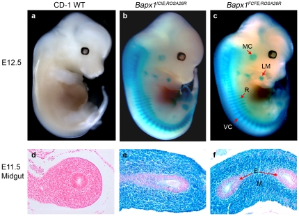
Figure 4. Functional analysis of Cre activity in the Bapx1ICIE;Rosa26R and Bapx1FCFE;Rosa26R mice.
(a–c) Whole-mount E12.5 Bapx1ICIE;Rosa26R and Bapx1FCFE;Rosa26R embryos stained with X-gal showing β-galactosidase activity in the correct anatomical context of Bapx1. (d–f) Transverse sections of E11.5 embryos confirmed X-gal stained cells in the mesenchymal cells of the midgut and not in the endothelium. MC, Meckel's cartilage; LM, limb mesenchyme; VC, vertebral column; R, ribs; M, mesenchyme; E, endothelium.

