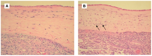
Figure 3. Histopathology of chorioamnion.
Hematoxylin and eosin stained histologic sections of chorioamnion (fetal membranes) are shown for a saline control (A) and GBS animal with chorioamnionitis (B). Neutrophils in the chorion are indicated with arrows in panel B, as well as being abundant in the decidua.

