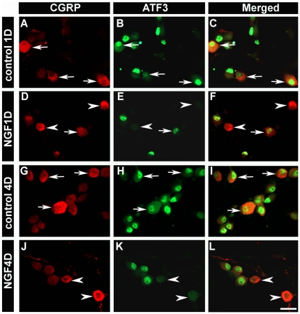
Figure 3. In vitro application of NGF decreased ATF3 expression in CGRP-positive neurons.
In control conditions (no growth factor), more than 90% of CGRP-positive neurons expressed ATF3 at 1 d (92.9±0.3%) (A–C; arrows) and 4 d (93.4±0.4%) (G–I; arrows) after plating. In NGF-treated cultures, the percentage of CGRP-positive neurons that expressed ATF3 was significantly decreased at 1 d (3.8±0.3%) (D–F; arrowheads) and 4 d (3.6±0.3%) (J–L; arrowheads). Scale bar = 50 µm. Two way ANOVA, p<0.01.

