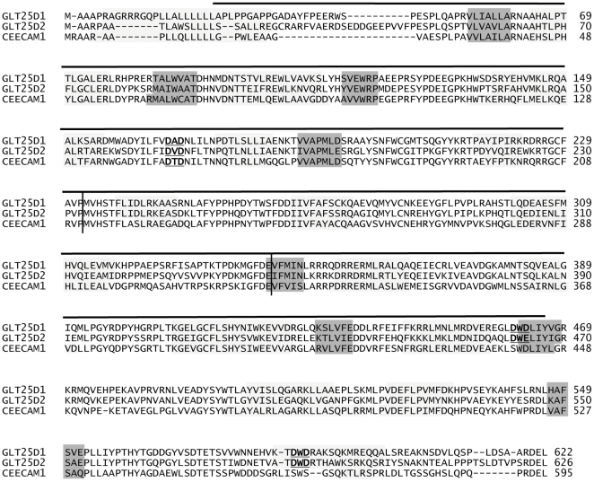
Figure 1. Structural prediction of GLT25D1, GLT25D2 and CEECAM1 proteins.
The protein sequences of human GLT25D1 (Swiss-Prot:Q8NBJ5), GLT25D2 (Swiss-Prot:Q81IYK4) and CEECAM1 (Swiss-Prot:Q5T4B2) were aligned using the PROMALS3D program. The sequences were analyzed and separated in two closely related groups, the first one with GLT25D1-D2 and the second one with CEECAM1, as determined with an identity threshold of 0.6. The secondary structure predictions are marked by shaded blocks, with light grey representing α-helices and dark grey β-strands. The predicted N-terminal, central and C-terminal domains are demarcated by vertical lines. The DXD motifs are underlined and marked in bold. The region ranging from amino acids 24 to 466 of human GLT25D1 structurally related to the Escherichia coli chondroitin polymerase (PDB ID: 2z86) is marked with a horizontal line above the sequences.

