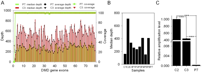
Figure 4. Detection of a deletion mutation of exon 1 in the DMD gene by targeted DNA-HiSeq.
(A) Diagram of the median depth, average depth and sequencing coverage for all 79 exons in the DMD gene. The median/average depth and the sequencing coverage for the first exon of the DMD gene in patient P7 (proband) was close to 0 and was different from the coverage at exon 1 in the control, C3 (proband's mother). (B) The median depth for exon 1 of the DMD gene in all of the 10 samples sequenced in one lane. The median depth for exon 1 was close to 0 in P7, but not in the other patients. (C) Verification of the deletion mutation in P7 by real-time PCR (mean ± SEM, ANOVA analysis). The results showed that the relative amplification (RA) level of exon 1 is nearly 0, and the RA level in C3 is approximately half of the RA level in C2 (p<0.001). Genomic β-actin DNA was used as a loading control.

