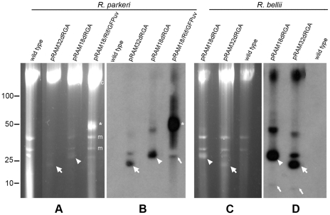
Figure 4. Plasmids in shuttle vector-transformed R. parkeri Oktibbeha and R. bellii RML369-C.
(A and C) A single PFGE gel divided into two panels showing the presence of plasmids in pRAM18dRGA and pRAM32dRGA- transformed R. parkeri and R. bellii. Full-length pRAM18/Rif/GFPuv–transformed R. parkeri is included for comparison. (B and D) Southern analysis of panels A and C respectively, hybridized with a digoxigenin-labeled gfpuv probe confirming the presence of plasmids pRAM18dRGA and pRAM32dRGA in transformed R. parkeri and R. bellii. Asterisks, arrowheads and wide arrows indicate the predominant putative supercoiled form of pRAM18/Rif/GFPuv, pRAM18dRGA and pRAM32dRGA respectively, and the narrow arrows indicate putative linear monomeric plasmid. Relative positions of ISE6 host cell mitochondrial, m, and chromosomal, c, DNAs are indicated to the right of panel A. Linear DNA marker positions in kbp are noted to the left of panel A.

