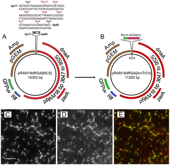
Figure 8. Transformation of R. montanensis with pRAM18dRGA[AmTrCh].
(A and B) Maps showing: (A) Restriction sites present in pRAM18dRGA[MCS]. (B) Insertion of AmTr/mCherry cassette into the MCS at the AvrII restriction site, yielding pRAM18dRGA[AmTrCh]. (C – E) Photomicrographs of cell free rickettsial suspensions centrifuged onto microscope slides and air dried showing: (C) GFPuv-positive R. montanensis transformed with pRAM18dRGA[AmTrCh], illuminated using the FITC filter. (D) Same microscopic field shown in C illuminated using the TRITC filter to reveal mCherry fluorescent positive rickettsiae. (E) Composite image made by merging images shown in C and D. Individual rickettsiae were examined on a TE2000-U Inverted microscope using epifluorescence illumination, with FITC and TRITC filter sets. Collected images of fluorescent rickettsiae from the red and green emission channels were processed in Image J. Bar = 10 µm.

