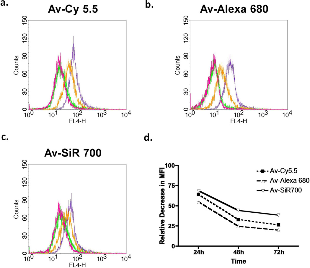
Figure 4.
Flow cytometry analysis of SHIN3 cells immediately (purple), 24 h (orange), 48 h (green), and 72 h (magenta) after PBS wash and removal of (a) Av-Cy5.5, (b) Av-Alexa 680, or (c) Av-SiR700 which demonstrates fluorescent signal degradation over time. Then, the amount of MFI degradation relative to T0 was compared (d). Av-SiR700 demonstrates less of a decrease in MFI from T0 than Av-Cy5.5 and Av-Alexa 680 at all time points.

