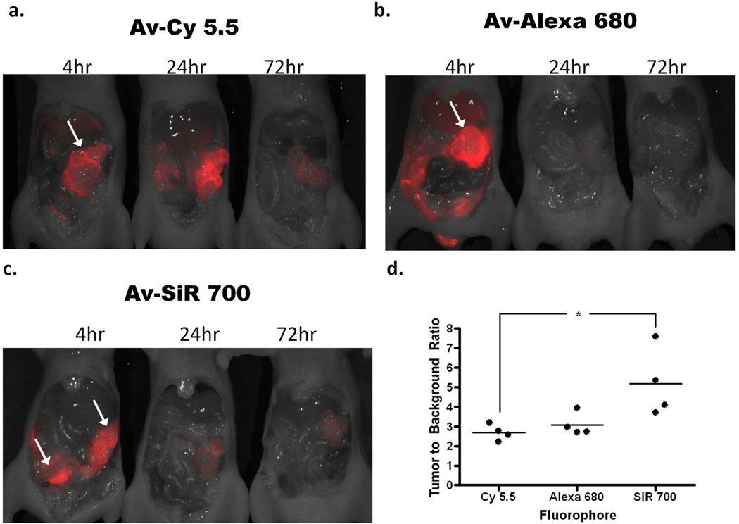
Figure 5.
In vivo fluorescence images of tumor-bearing mice 4, 24, or 72 h after intraperitoneal injection of either (a) Av-Cy5.5, (b) Av-Alexa 680, or (c) Av-SiR700. Images clearly identify SHIN3 tumors within the abdomen (arrows). Images clearly demonstrate the degradation of each NIR imaging probe over time. Interestingly, Av-Cy5.5 appears to demonstrate an increase in fluorescence intensity of tumor from 4 to 24 h after Av-Cy5.5 injection. Whole body in vivo images were then used to calculate the TBR for each NIR imaging probe at 4 h after injection. ROIs were drawn around the tumors and ROIs of the same size were drawn over the adjacent abdomen to determine background signal. This is represented graphically (d), demonstrating that Av-SiR700 has a relatively higher TBR than Av-Alexa 680, but Av-SiR700 has a significantly greater TBR than Av-Cy5.5, (p<0.05).

