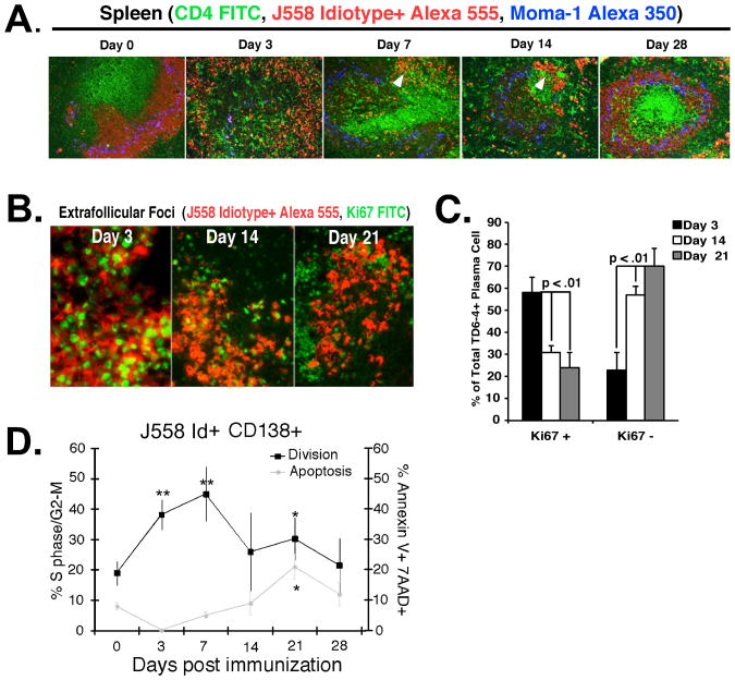
Figure 3. Extrafollicular J558 Id+ CD138+ early proliferation is balanced by apoptosis later in the response to E. cloacae.
(A) Spleen sections from VHJ558 TG mice immunized with E. cloacae strain MK7 were harvested at 0, 3, 7, 14, and 28 days after immunization and stained with antibodies specific for DEX-specific B cells bearing the J558 idiotype (red), CD4+ T cells (green), and metallophilic macrophages (blue) revealing the presence of J558 idiotype high plasma cells within extrafollicular foci in the red pulp (white arrows). (B) Many more extrafollicular J558 Id+ CD138+ B cells express Ki67 at 3 days compared to 21 days after immunization with E. cloacae. Sections of spleen from VHJ558 TG mice immunized with E. cloacae strain MK7 and stained with Ki67-FITC (green) and anti-J558 Id (red). (C) Ki67+ J558 Id+ plasmablasts in extrafollicular foci at 3, 14, and 21 days after E. cloacae strain MK7 immunization. Numbers were calculated by counting Ki67 staining cells in 5 individual extrafollicular foci per 3 mice. (D) DEX-specific plasmablast homeostasis is balanced by proliferation and apoptosis. Analysis of division and apoptosis of J558 Id+ CD138 + B cells using intracellular PI cell to determine cycle status (black line; left y axis) and surface annexin V+/7AAD+ staining to determine late apoptotic cells (grey line; right y axis). Data shown is from 3 independent experiments (n= 3 mice per experiment). Asterisks show statistical significance in comparison to the day 0 time point.

