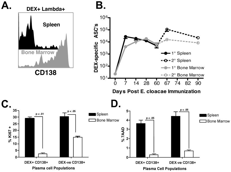
Figure 4. A quiescent population of DEX+ λ+ CD138+ ASCs accumulate in the bone marrow and display increased intracellular immunoglobulin expression.
(A) The expression of CD138 on surface positive DEX+ λ+ B cells in the spleen (black histogram) and bone marrow (grey histogram) 60 days after E. cloacae strain MK7 immunization. Data is representative of 5 mice. (B) DEX-specific λ+ ASC numbers were measured in the spleen after primary (black line, closed circles, E. cloacae strain MK7 injected on day 0) and secondary (dashed black line, open circles, E. cloacae strain MK7 re-challenge on day 60) and bone marrow after primary (grey line, closed circles, E. cloacae strain MK7 injected on day 0) and secondary (dashed grey line, open circles, E. cloacae strain MK7 injected on day 60) immunization; n= 5 mice per time point. (C) Proportions of Ki67 positive DEX+ λ+ CD138+ and DEX negative CD138+ B cells in the spleen (black bars) and bone marrow (white bars) 60 days post E. cloacae strain MK7 immunized BALB/c mice. (D) Proportions of 7AAD positive DEX+ λ+ CD138+ and DEX negative CD138+ B cells in the spleen (black bars) and bone marrow (white bars) in 60 days post E. cloacae strain MK7 immunized BALB/c mice. Proportions shown for (C) and (D) were calculated from 5 mice.

