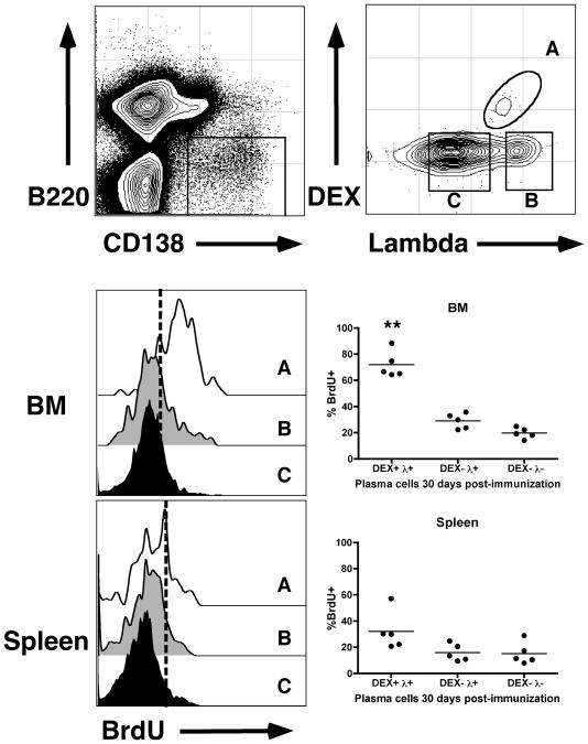
Figure 5. DEX-specific ASCs in the bone marrow are long-lived.
Immunized BALB/c mice were fed BrdU in drinking water for 7 days after injection. 4 weeks after injection, mice were sacrificed and levels of BrdU incorporation were determined. Top panel shows the electronic gates used to determine DEX-specific ASCs (Gate A), DEX negative Lambda light chain + ASCs (Gate B) and DEX and Lambda light chain double negative ASCs (Gate C). Middle and lower panels show representative histograms and percent BrdU staining positively in each of the three populations A, B and C in the bone marrow and spleen respectively. Dashed lines in the histograms determine background BrdU staining based on negative controls. Asterisks show statistical significance in comparison to DEX+ λ+ ASCs in the spleen.

