
Figure 6. DEX is associated with MZ macrophages early and later with CD11c+ dendritic cells and J558 Id+ plasma cells at extrafollicular sites after I.V. injection of BALB/c mice.
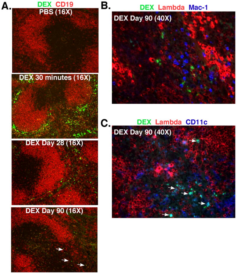
(A) DEX (green) is located in the splenic MZ containing CD19+ B cells (red) at 30 minutes and later at 28-90 days DEX is found predominantly in the red pulp. White arrows indicate the presence of DEX in BALB/c mice 90 days post injection. (B+ C) DEX (green) does not associate with Mac-1+ neutrophils (blue) distinguished by their multi-lobed nuclei, but associates with CD11c+ DCs (blue/blue green) and λ+plasma cells (bright red) in BALB/c mice at 90 days after immunization with DEX. White arrows indicate the association of DEX (green) with CD11c+ dendritic cells (blue). Each image shown is representative of 3 mice per time point.