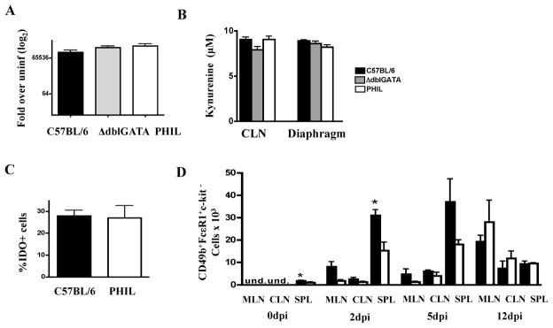
Fig. 3. IDO response and basophilia in eosinophil-ablated mice.
(A) IDO gene expression in diaphragms of infected ΔdblGATA, PHIL, and WT mice 15dpi.(B) KYN in cultures of CLN and diaphragm leukocytes collected 15dpi. (C) Detection of IDO indiaphragm leukocytes from infected WT and PHIL mice collected 12dpi. (D) Basophils in WT and ΔdblGATA lymphoid tissues. Basophils were identified as CD49b+FcεR1+c-kit− cells in the CLN, MLN, and spleen (SPL) in uninfected and infected mice 2, 5, and 12 dpi by flow cytometry. Experiments were performed two times with similar results. Values represent means +/− SD, n = 3 – 4 mice. Significant differences were determined by Student’s t test. *p<0.05.

