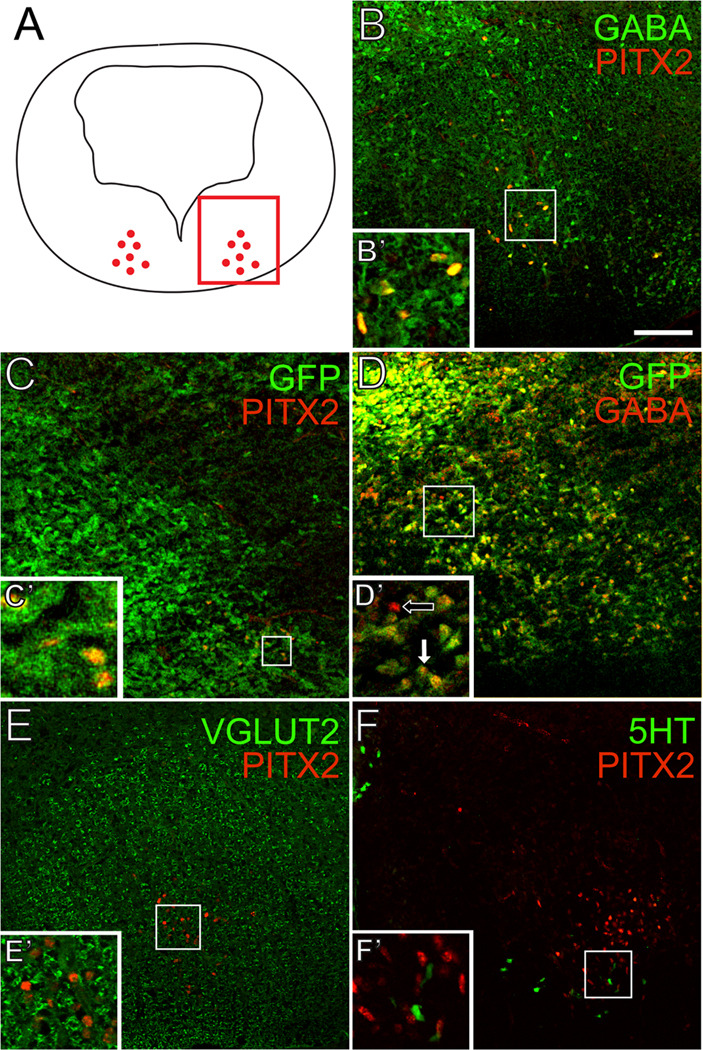
Figure 2. PITX2-positive GABAergic neurons in r1 are distinct from serotonergic, glutamatergic, and cholinergic neurons.
Double immunofluorescence of E14.5 mouse brain tissues sectioned transversely at the level of r1. Schematic in A indicates transverse orientation for panels B–F. Square inset in A shows the area represented in B–F. PITX2 co-localizes with GABA (B–B’) and GAD67-GFP (C–C’). Most GFP-positive cells are also GABA-positive (D–D’). PITX2-positive cells are negative for VGLUT2 (E–E’) and 5-HT (F–F’). Scale bar in B is 100 µm and applies to panels B–F. All images were taken using confocal microscopy.

