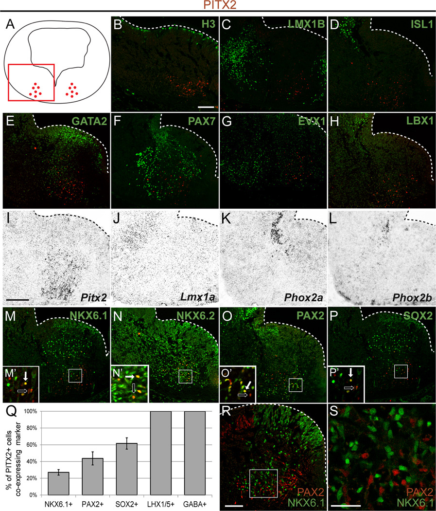
Figure 4. PITX2-positive GABAergic neurons occupy distinct regions of the ventral hindbrain.
Double immunolabeling (A–H, M–P, R, S) or in situ hybridization (I–L) of E12.5 mouse brain tissues sectioned transversely at the level of r1 (orientation as in panel A) reveals distinct patterns of overlap between PITX2 and several transcription factors. Insets in M–P are enlarged in M’–P’, and show double (solid arrow) or single (open arrow) labeled PITX2-positive cells. Graph in Q indicates the percentage of PITX2-positive cells which co-express the marker indicated (NKX6.1, PAX2, SOX2, and LHX1/5 are from E12.5 embryos; GABA is from E14.5 embryos). Error bars are +/− standard error of the mean of cell counts from N≥3 sections. (R, S) PAX2 and NKX6.1 mark separate cell populations in r1. Cells in the intermediate layer of panel R are enlarged in S. Scale bar in B is 100 µm and applies to panels B–H and M–P. Scale bar in I is 100 µm and applies to panels I–L. Scale bars in R and S are 75 µm and 30 µm, respectively. All immunofluorescent images were taken using confocal microscopy.

